
Research · Injury Prevention
The Weakest Link: What the Evidence Says About Growth Plate Injury Risk in Young Athletes
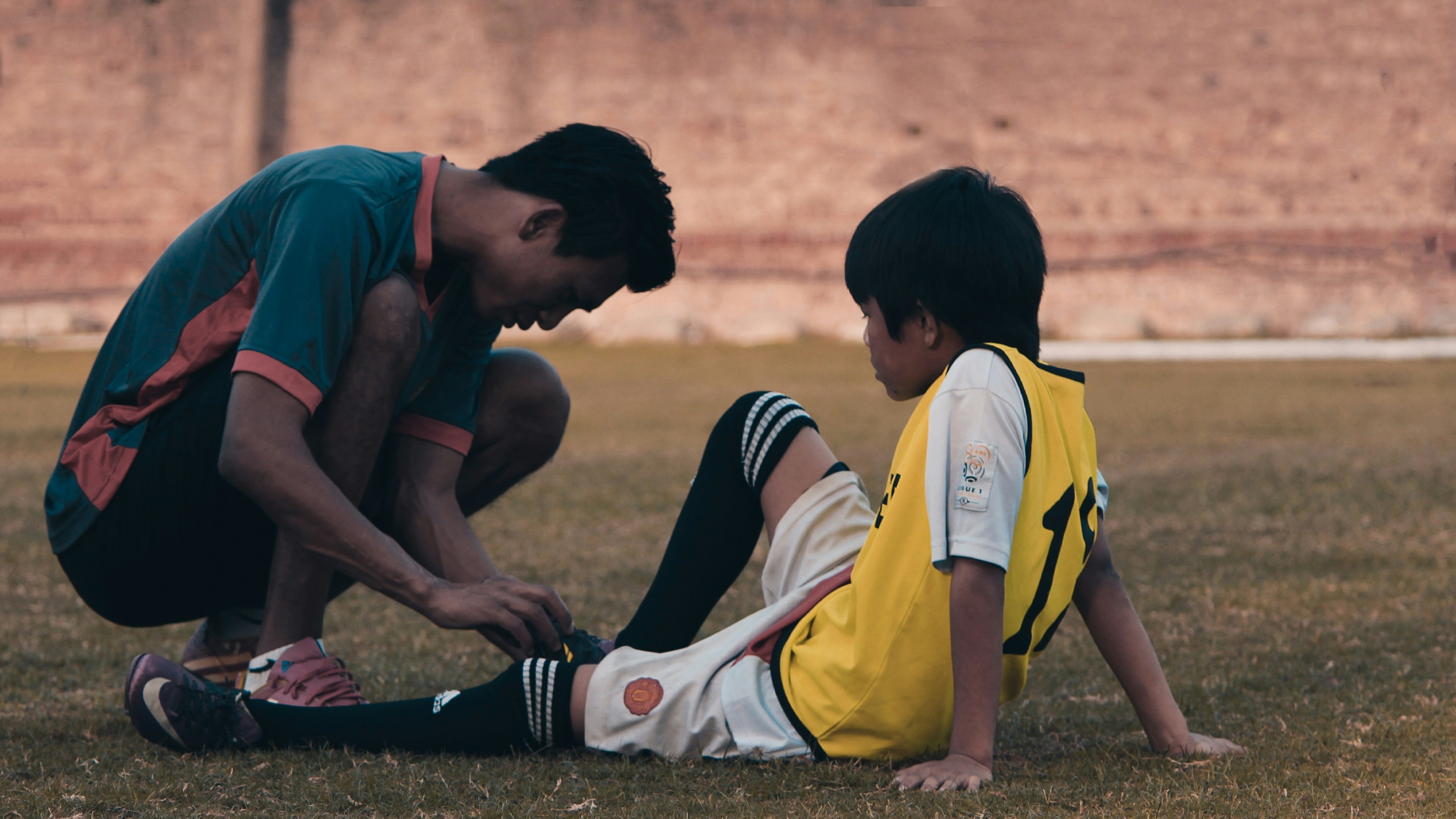
"Watch the growth plates" is one of the more common instructions handed to parents and coaches of a young athlete, and one of the least specific. A growth plate is a real, identifiable structure — not a euphemism for general caution — with a documented mechanical reason for being more fragile at specific points in development, and a short list of specific, well-studied conditions that show up when that mechanism gets overloaded. The mechanistic version of the concept changes what "watching" it should actually mean in a training plan.
The growth plate, formally the physis, is a layer of cartilage near the end of a long bone responsible for the bone's lengthwise growth until it ossifies into solid bone at skeletal maturity.1 A related structure, the apophysis, is a secondary growth centre where a major tendon anchors onto bone rather than where two bones articulate at a joint — the tibial tuberosity below the kneecap, the back of the heel bone, the inside of the elbow, the top of the shoulder. Nearly every overuse condition specific to growing athletes traces back to one of these apophyseal sites, which is why they are the focus here rather than the joint-line growth plates more commonly associated with an acute fracture.
Why it is structurally the weakest point
Skeletal tissue in a child does not fail under load the way it does in an adult, and the difference is structural rather than a general statement about fragility. A pediatric orthopaedic reference on physeal injury states that the growth plate is "five times more brittle than the surrounding ligaments," and that in a child the physis is weaker than the adjacent ligaments or periosteum — so a force that would sprain a ligament or strain a tendon in an adult is more likely to injure the growth plate first in a child or adolescent.1 That relationship reverses once the skeleton fully matures, which is the specific reason growth-plate injury is a paediatric and adolescent phenomenon rather than a lifelong one.
What actually changes during a growth spurt
That baseline vulnerability is not fixed across childhood — it rises sharply during the adolescent growth spurt itself, the period researchers refer to as peak height velocity (PHV), the point of fastest growth in standing height during puberty. A systematic review of physeal injury in youth sport found that "the susceptibility of the growth plate to injury appears to be especially pronounced during periods of rapid growth," attributing this to structural change in the plate itself: an increase in growth rate is "accompanied by structural changes that result in a thicker and more fragile plate."2 The same review flags a second, compounding factor — bone mineralisation can lag behind bone lengthening during the pubertal growth spurt, leaving the bone "temporarily more porous" at precisely the point it needs to support more force from a taller, heavier frame.2
This is not a theoretical concern read off a mechanism diagram — it shows up directly in injury data. A three-year study following 26 talented youth soccer players longitudinally through their individual growth spurts found significantly more traumatic injuries in the year of PHV itself (1.41 per player) than in the year before it (0.81), and a comparable rise in overuse injuries across the same before-to-after window (0.81 to 1.41 per player per year). Days missed to injury climbed from 7.27 to 15.69 per player per year during the PHV year.3 The authors' own conclusion is direct: the adolescent growth spurt increases vulnerability to traumatic injury while it is happening, and to overuse injury in its immediate aftermath.3 Because PHV timing varies by roughly two years between individual athletes of identical chronological age, a squad organised strictly by birth year will always contain some athletes moving through this higher-risk window and others who are nowhere near it — a detail with direct implications for how load gets assigned across that group.
The mechanism behind the specific conditions
The conditions below share a single underlying mechanism. During a growth spurt, the bone itself lengthens faster than the muscle-tendon unit crossing it can adapt, leaving that muscle and tendon relatively short and tight for the new length of lever they now cross. The tension this creates concentrates at the one point built to move rather than resist it — the apophysis, where the tendon actually anchors to bone. A clinical reference on the most common of these conditions puts it directly: "bone growth exceeds the ability of the muscle-tendon unit to stretch sufficiently to maintain previous flexibility," and because the apophysis is the weakest link in that entire muscle-tendon-bone chain, it is where repetitive loading concentrates its damage first.4 Different sports load different apophyses, which is why these conditions cluster in specific, sport-linked patterns rather than appearing at random.
Osgood-Schlatter disease
Osgood-Schlatter disease is irritation, and in more advanced cases partial avulsion, of the apophysis at the tibial tuberosity, where the patellar tendon attaches below the kneecap — driven by repetitive quadriceps loading through jumping, sprinting, and kicking.4 Its onset tracks growth timing closely, coinciding with each sex's own adolescent growth spurt: typically ages 10 to 15 in boys and 8 to 13 in girls.4 A retrospective study of adolescent athletes found the condition presenting bilaterally in a meaningful share of cases, and resolving in the great majority within about a year with load modification rather than surgery.5 Population-level data puts prevalence in 12-to-15-year-olds at roughly 9.8% overall — 11.4% in boys against 8.3% in girls — with prevalence in sports-active adolescents specifically running meaningfully higher than that.4 Football and basketball show up disproportionately in the sport breakdown, tracking the jumping and kicking demand the condition is mechanically tied to.4
Sever's disease
Sever's disease, or calcaneal apophysitis, is the equivalent condition at the back of the heel, where the Achilles tendon anchors onto the growing calcaneus. It is the most common cause of heel pain in children and young adolescents, typically presenting between ages 8 and 15, with average age at diagnosis around 11 to 12.6 Boys are affected two to three times more often than girls, and up to 60% of cases present in both heels at once — a pattern consistent with a systemic growth-timing driver rather than a one-off local injury.6 High-impact sports with repeated ground contact — soccer, track and cross-country, gymnastics — are the consistent common thread, and clinical guidance links onset directly to a recent growth spurt, a new sport, or the start of a season, rather than to any single traumatic event.6
Little League shoulder and elbow
Overhead throwing loads two growth-plate sites specifically, and both carry the sport's name because youth baseball pitching is where the mechanism is most studied. Little League elbow — medial epicondyle apophysitis — is traction stress on the growth plate at the inside of the elbow, generated by the valgus, inward-bending load of the throwing motion. In a young thrower, that open apophysis is roughly five times weaker than the adjacent ulnar collateral ligament, so the growth plate gives way before the ligament does.7 Radiographic screening of competitors at Little League regional and national championships found medial epicondyle displacement in 57% of those X-rayed, and prevalence peaks at around 30% among 11-to-12-year-old throwers specifically.7
Little League shoulder — proximal humeral epiphysiolysis — is the same overload pattern at the growth plate near the top of the humerus. A biomechanical study of 14 elite youth pitchers, filmed throwing fastballs under game-simulated conditions, found that the shear stress generated late in the arm-cocking phase of a throw is large enough to deform the still-cartilaginous proximal humeral growth plate directly.8 Because that plate contributes roughly 80% of the humerus's total lengthwise growth, it stays mechanically vulnerable well into mid-adolescence, and the resulting condition is seen almost exclusively in overhead-throwing athletes aged 11 to 16.9
The clearest evidence tying throwing volume itself to serious injury risk comes from a ten-year prospective study of 481 youth pitchers aged 9 to 14, followed from 1999 to 2008, using elbow surgery, shoulder surgery, or retirement due to a throwing injury as its injury definition. Pitchers who threw more than 100 innings in a single year were 3.5 times more likely to sustain one of those serious injuries than pitchers who did not.10 That is a dose-response relationship reported in the sport's own primary literature, not an inference — innings actually pitched, not talent or mechanics alone, moved the injury rate.
The growth plate does not fail because a young athlete trained too hard in some general sense. It fails because the loading pattern stayed the same while the tissue underneath it did not.
Evidence-based prevention and monitoring
None of this is an argument for pulling a young athlete out of sport during a growth spurt. It is an argument for specific, testable adjustments to how load is assigned and tracked while a growth spurt is actually happening.
The clearest working example already exists in baseball. Pitch Smart, the joint guideline programme from Major League Baseball and USA Baseball developed with input from the American Sports Medicine Institute, sets age-scaled daily pitch-count ceilings — roughly 50 pitches at ages 7–8, rising through the mid-70s to mid-80s by ages 11–12 — paired with mandatory rest days that scale with how many pitches were actually thrown, restrictions on breaking pitches before the fastball and changeup are established, and a standing rule against pitching in games on three consecutive days regardless of pitch count.11 It is a direct, structural answer to the innings-based injury-risk finding above: cap the dose and mandate the recovery, rather than leaving either to judgement in the moment.
The second piece is identifying the higher-risk window itself, rather than assuming it from age. Because PHV timing varies so much between individual athletes, a validated, non-invasive method exists for estimating how close an athlete actually is to their own growth spurt: the maturity offset equation, derived from standing height, sitting height, estimated leg length, body mass, and chronological age, predicting years to or from peak height velocity without needing an X-ray.12 Tracking height at regular intervals — the systematic review cited above specifically recommends measuring every three months — turns "somewhere in the growth spurt" from a guess into a measured, trackable value, which is what actually allows a program to reduce load and delay technical progressions for the specific athletes moving through that window, rather than applying that caution to everyone or no one.2
The third piece is treating maturity, not birth year, as the variable that should drive individual programme design. The NSCA's own position statement on long-term athletic development is explicit that two athletes of identical chronological age can be years apart in biological maturity, and that programming built around age rather than developmental stage is measuring the wrong thing.13 Applied to growth-plate risk specifically, that means the athlete who has just entered a growth spurt needs a different load, technical emphasis, and monitoring frequency than a teammate of the same age who has not — a distinction a calendar-based programme has no way to make on its own.
Put together, the practical checklist is short:
- Track height, not just age. A measurable change in growth rate is the actual signal that a young athlete has entered the higher-risk window — not a birthday.
- Treat pain that tracks with activity as data, not drama. Pain that worsens during or after training and eases with rest is the consistent early pattern across Osgood-Schlatter, Sever's, and Little League shoulder and elbow alike — a different pattern from pain with no relationship to activity at all.
- Cap and rotate the load at known high-risk sites. Age-scaled pitch counts with mandatory rest are the clearest existing model; the same logic — a volume ceiling paired with enforced recovery — applies to any single, repetitive movement pattern loading one apophysis over and over across a season.
The evidence points to the same conclusion from three separate directions — the structural biology of the physis itself, the injury data gathered around peak height velocity, and the sport-specific volume studies in throwing athletes. A growth plate is not, on its own, a reason to train less. It is a specific, identifiable, temporarily vulnerable structure that a well-designed programme can actually plan around, once its status is being measured rather than assumed.
Sources
- Waseem M, Taqi M, Marquart MJ. "Pediatric Physeal Injuries Overview." In: StatPearls [Internet]. Treasure Island (FL): StatPearls Publishing. NCBI Bookshelf: NBK560546.
- Caine D, DiFiori J, Maffulli N. "Physeal injuries in children's and youth sports: reasons for concern?" British Journal of Sports Medicine 40(9):749–760, 2006. Full text (PMC).
- van der Sluis A, Elferink-Gemser MT, Coelho-e-Silva MJ, Nijboer JA, Brink MS, Visscher C. "Sport Injuries Aligned to Peak Height Velocity in Talented Pubertal Soccer Players." International Journal of Sports Medicine 35(4):351–355, 2014. DOI: 10.1055/s-0033-1349874.
- Smith JM, Varacallo MA. "Osgood-Schlatter Disease." In: StatPearls [Internet]. Treasure Island (FL): StatPearls Publishing. NCBI Bookshelf: NBK441995.
- Kujala UM, Kvist M, Heinonen O. "Osgood-Schlatter's Disease in Adolescent Athletes: Retrospective Study of Incidence and Duration." The American Journal of Sports Medicine 13(4):236–241, 1985. DOI: 10.1177/036354658501300404.
- Smith JM, Varacallo MA. "Sever Disease (Calcaneal Apophysitis)." In: StatPearls [Internet]. Treasure Island (FL): StatPearls Publishing. NCBI Bookshelf: NBK441928.
- Hodge C, Schroeder JD. "Medial Epicondyle Apophysitis (Little League Elbow)." In: StatPearls [Internet]. Treasure Island (FL): StatPearls Publishing. NCBI Bookshelf: NBK570592.
- Sabick MB, Kim YK, Torry MR, Keirns MA, Hawkins RJ. "Biomechanics of the Shoulder in Youth Baseball Pitchers: Implications for the Development of Proximal Humeral Epiphysiolysis and Humeral Retrotorsion." The American Journal of Sports Medicine 33(11):1716–1722, 2005. DOI: 10.1177/0363546505275347.
- Collins AC, May T. "Proximal Humeral Epiphysiolysis." In: StatPearls [Internet]. Treasure Island (FL): StatPearls Publishing. NCBI Bookshelf: NBK534301.
- Fleisig GS, Andrews JR, Cutter GR, Weber A, Loftice J, McMichael C, Hassell N, Lyman S. "Risk of Serious Injury for Young Baseball Pitchers: A 10-Year Prospective Study." The American Journal of Sports Medicine 39(2):253–257, 2011. DOI: 10.1177/0363546510384224.
- Major League Baseball & USA Baseball, developed with the American Sports Medicine Institute. "Pitch Smart: Pitching Guidelines." pitchsmart.org.
- Mirwald RL, Baxter-Jones ADG, Bailey DA, Beunen GP. "An Assessment of Maturity from Anthropometric Measurements." Medicine & Science in Sports & Exercise 34(4):689–694, 2002. DOI: 10.1097/00005768-200204000-00020.
- Lloyd RS, Cronin JB, Faigenbaum AD, Haff GG, Howard R, Kraemer WJ, Micheli LJ, Myer GD, Oliver JL. "National Strength and Conditioning Association Position Statement on Long-Term Athletic Development." Journal of Strength and Conditioning Research 30(6):1491–1509, 2016. Full position statement (PDF, NSCA).
Want this applied to your own program, team, or school? Free 20-minute consult, no obligation.
Book a free consult →